Resurrected Ancient Enzyme Offers New Window Into Early Earth And The Search For Life Beyond It
By resurrecting a 3.2-billion-year-old enzyme and studying it inside living microbes, researchers at the University of Wisconsin–Madison have created a new way to improve our understanding of the origins of life on Earth and possibly recognize signs of life elsewhere.
Recently published in Nature Communications, the NASA-funded study uses synthetic biology to reverse-engineer modern enzymes and rebuild their possible ancestors. Betül Kaçar, a professor of bacteriology, and Holly Rucker, a PhD candidate in Kaçar’s lab, focused on an enzyme called nitrogenase, which is critical to the process that converts atmospheric nitrogen into a form usable by living organisms. “We picked an enzyme that really set the tone of life on this planet and then interrogated its history,” Kaçar says. “Without nitrogenase, there would be no life as we know it.”
Historically scientists have relied on evidence found in the geological record to build our understanding of past life on Earth. Such significant fossil and rock samples are and often require a bit of luck to find. Kaçar and Rucker see synthetic biology as a way to augment this important work, filling in the gaps by creating tangible reconstructions of ancient enzymes, putting them into microbes, and studying them in a modern lab. “Three billion years ago is a vastly different Earth than what we see today,” says Rucker.
Back before the Great Oxidation Event, she explains, the atmosphere contained more carbon dioxide and methane, and life primarily consisted of anaerobic microbes. Being able to understand how these microbes accessed a nutrient as vital as nitrogen offers a sharper picture of how life persisted and evolved in the window of time before oxygen-dependent organisms began reshaping the planet.
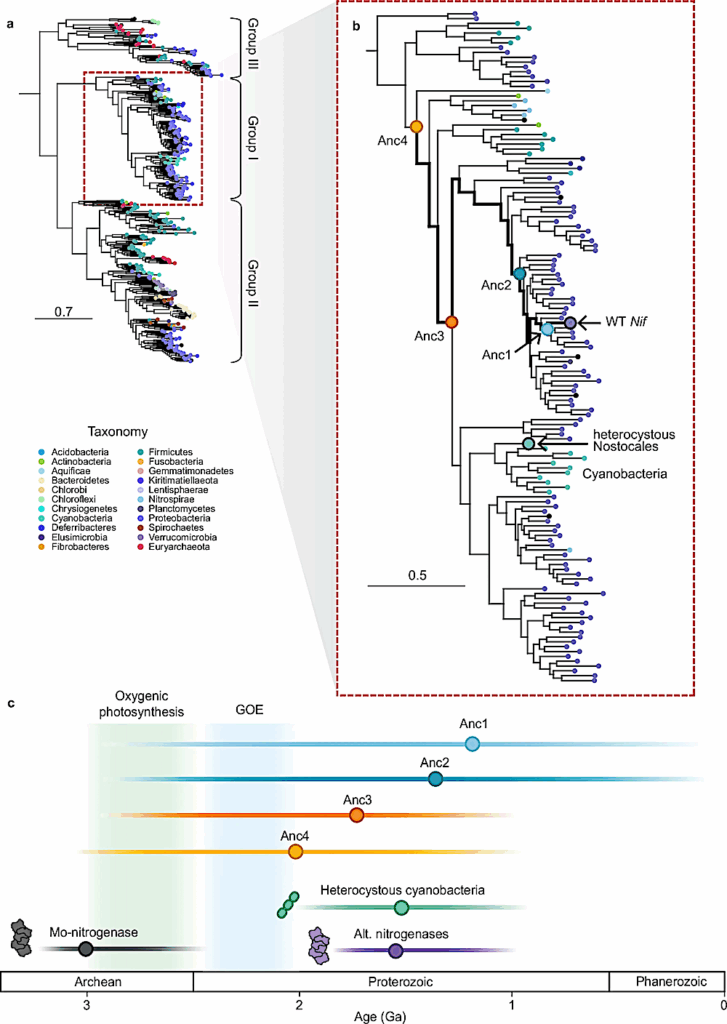
a Node colors represent the extant taxonomy. b Close-up of Group I nitrogenases. The ancestral nodes targeted in this study are labeled with circles. Heterocystous cyanobacteria (highlighted in teal) serve as a minimum age constraint for the ancestral nitrogenase nodes based on fossil evidence32. c Approximate ages for the origin of nitrogenase isoforms are based on geochemical12 and phylogenetic data29. The estimated timing of the origin of oxygenic photosynthesis and the Great Oxidation Event (GOE) are shaded in green and blue, respectively. — Nature
While there are not fossilized enzymes the team can study, these enzymes can leave behind recognizable signatures in the form of isotopes, which researchers can measure in rock samples. But much of that work relied on the assumption that ancient enzymes produce the same isotopic signatures as modern versions. Rucker began to wonder: Are we actually interpreting the rock record correctly?
“It turns out, yes, at least for nitrogenase,” Rucker says. “The signatures that we see in the ancient past are the same that we see today, which then also tells us more about the enzyme itself.” The team found that even though ancient nitrogenase enzymes have different DNA sequences than modern versions, the mechanism that controls the isotopic signature preserved in the rock record has stayed the same. Rucker hopes to investigate why the mechanism was conserved while other aspects of the enzyme evolved.
This project connects to Kaçar’s broader work as the leader of MUSE, a NASA-funded astrobiology research consortium based at UW–Madison. From astrobiologists to geologists across several institutions, MUSE brings researchers together to strengthen NASA space missions through new evolutionary insights into microbiology and molecular biology on Earth.
With nitrogenase-derived isotopes now identified as a reliable biosignature on Earth, MUSE has a clearer framework for evaluating similar signals that may be found on other planets. “As astrobiologists, we rely on understanding our planet to understand life in the universe. The search for life starts here at home, and our home is 4 billion years old,” Kaçar says. “So, we need to understand our own past. We need to understand life before us, if we want to understand life ahead of us and life elsewhere.”
Resurrected nitrogenases recapitulate canonical N-isotope biosignatures over two billion years, Nature
Astrobiology, Genomics, evolution,