Specific Role Of Two NlpC/P60 Endopeptidases In Cell Division And Membrane Vesicle Formation In Deinococcus radiodurans
The bacterial cell wall is composed of peptidoglycan (PG), a sugar polymer cross-linked by short peptide stems. PG determines cell morphology and protects it from environmental stresses.
Cell growth and division require a balance between synthesis and hydrolysis of the PG. One class of PG hydrolase is the NlpC/P60 superfamily which is broadly distributed in bacteria, archaea, eukaryotes and viruses. Deinococcus radiodurans, characterized by its extreme radioresistance, is a diderm bacterium with a thick layer of PG located between the inner and outer membrane. D. radiodurans exhibits three NlpC/P60 endopeptidases, their role in morphogenesis and cell cycle remains unexplored.
In this work, we investigated the role of each endopeptidase in cell division to assess their specific role. Here, we showed that the CwlB protein is involved in cell division and that cwlA gene is essential for cell viability. The CwlC protein is not required for cell shape maintenance or cell division. We showed that CwlA protein is homogeneously localized around the cell except on septal region.
CwlA-depleted cells lost viability, displayed morphological changes, and produced numerous membrane vesicles, similarly to cells exposed to sublethal mitomycin C (a DNA-damaging agent) or to DdrO depletion, the transcriptional repressor of the main genotoxic stress response in Deinococcus. We showed that cwlA expression is highly repressed under DdrO depletion. These data suggested a link between response pathway to genotoxic conditions and cell wall remodeling.
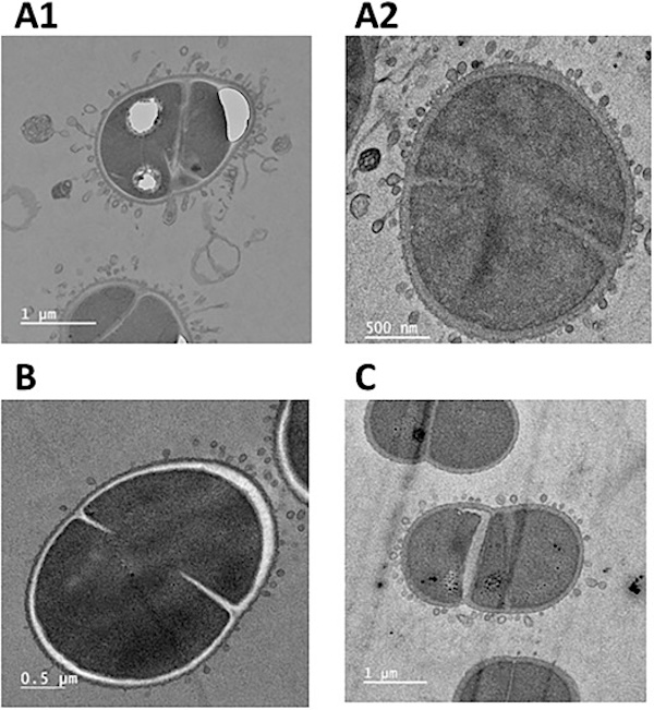
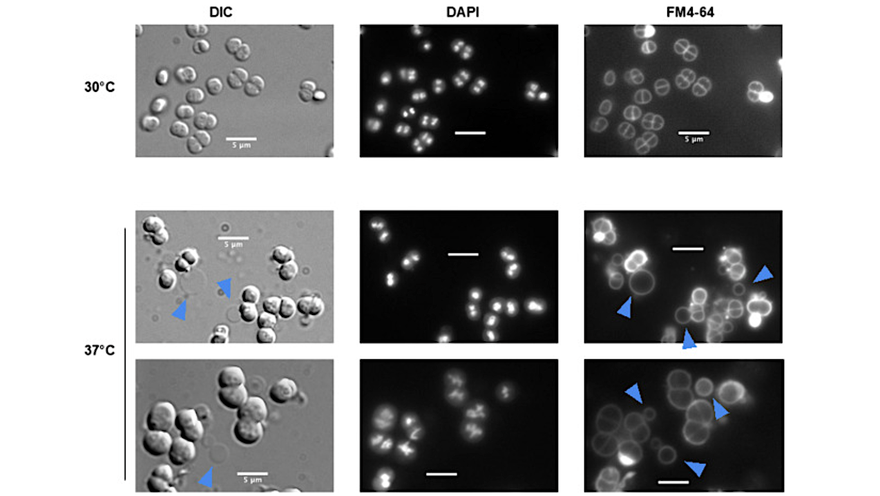
Transmission electron microscopy photograph of membrane vesicles in D. radiodurans. Impact of CwlA depletion on D. radiodurans cells. A and C: Cells in exponential growth phase cultivated at 30 °C in medium supplemented with spectinomycin were harvested by centrifugation, diluted in antibiotic-free medium and incubated at 37 °C for 24 h. A1 and A2 -GY 18249 (ΔcwlA/ prepUTS::cwlA+); C – GY14164 (ΔddrO/prepUTS::ddrO+). B: Membrane vesicle formation in wild type cells in the presence of mitomycin C. — Current Research in Microbial Sciences
- Specific role of two NlpC/P60 endopeptidases in cell division and membrane vesicle formation in Deinococcus radiodurans, Current Research in Microbial Sciences via PubMed
- Specific role of two NlpC/P60 endopeptidases in cell division and membrane vesicle formation in Deinococcus radiodurans, Current Research in Microbial Sciences
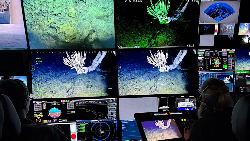
Astrobiology, extremophile,