Tricorder Tech: A Non-Invasive, Label-Free Method for Examining Tardigrade Anatomy Using Holotomography
Background/Objectives: Holotomography is an advanced imaging technique that enables high-resolution, three-dimensional visualization of microscopic specimens without the need for fixation or staining.
Here we aim to apply holotomography technology to image live Hypsibius exemplaris in their native state, avoiding invasive sample preparation procedures and phototoxic effects associated with other imaging modalities.
Methods: We use a low concentration of 7% ethanol for tardigrade sedation and sample preparation. Holotomographic images were obtained and reconstructed using the Tomocube HT-X1 system, enabling high-resolution visualization of tardigrade anatomical structures.
Results: We captured detailed, label-free holotomography images of both external and internal structures of tardigrade, including the digestive tract, brain, ovary, claws, salivary glands, and musculature.
Conclusions: Our findings highlight holotomography as a complementary high-resolution imaging modality that effectively addresses the challenges faced with traditional imaging techniques in tardigrade research.
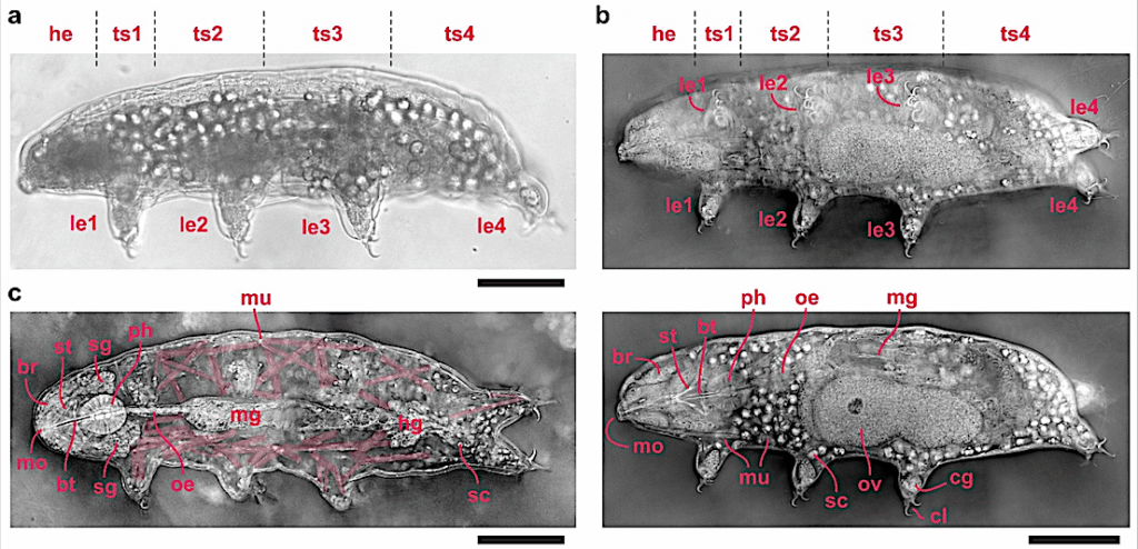
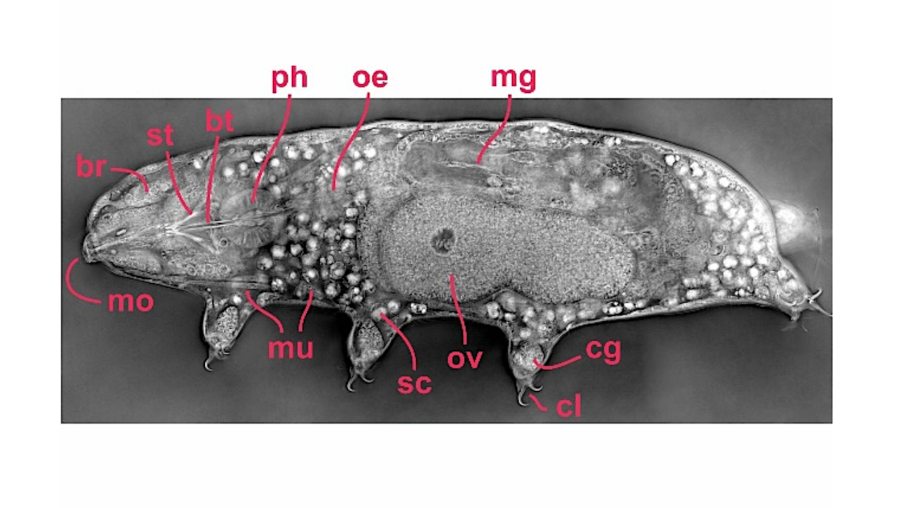
Anatomy of tardigrade visualized using holotomography. (a) Brightfield image of a tardigrade. (b) Reconstructed holotomogram at two different z-planes, with the upper panel highlighting the external morphology and the lower panel revealing internal anatomy, including the digestive tract, ovary, and others. (c) Reconstructed holotomogram illustrating musculature, highlighted in light red. Scale bar: 50 µm. Abbreviations: he, head; ts1–4, trunk segments 1–4; le1–4, legs 1–4; br—brain; st—stylet; bt—buccal tube; ph—pharynx; oe—oesophagus; mg—midgut; mg—hindgut; mo—mouth; mu—muscle; sc—storage cells; sg—saliva glands; ov—ovary; cl—claws; cg—claw glands. — Tomography, via PubMed
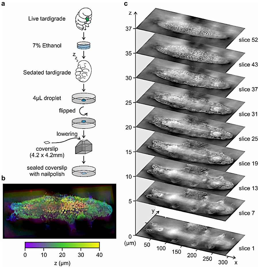
Tardigrade 3D holotomography. (a) Schematic of tardigrade preparation for holotomography imaging. Tardigrades are sedated in 7% ethanol in tap water and placed in a 4 µL droplet at the center of a 6-well plate. A 4.2 × 4.2 mm coverslip is carefully positioned by lowering the plate until the droplet comes into contact with and adheres to the coverslip. The coverslip is sealed with nail polish to prevent evaporation during imaging. (b) Three-dimensional reconstruction derived from the holotomography, presented with a pseudo-gradient color scale (purple–green–yellow) to indicate image depth (0 μm–20 μm–40 μm). (c). Representative slices from the reconstructed holotomography stack, showcasing a z-resolution of 714 nm and an xy-resolution of 156 nm (Table S1).– Tomography, via PubMed
- Tricorder Tech: A Non-Invasive, Label-Free Method for Examining Tardigrade Anatomy Using HolotomographyTomography, via PubMed (open access)
- A Non-Invasive, Label-Free Method for Examining Tardigrade Anatomy Using Holotomography, Tomography,(open access)
Astrobiology, Astrochemistry,