Reduced Pseudomonas Aeruginosa Cell Size Observed on Planktonic Cultures Grown in the International Space Station
Bacterial growth and behavior have been studied in microgravity in the past, but little focus has been directed to cell size despite its impact on a myriad of processes, including biofilm formation, which is impactful regarding crew health.
To interrogate this characteristic, supernatant aliquots of P. aeruginosa cultured on different materials and media on board the International Space Station (ISS) as part of the Space Biofilms Project were analyzed. For that experiment, P. aeruginosa was grown in microgravity—with matching Earth controls—in modified artificial urine medium (mAUMg-high Pi) or LB Lennox supplemented with KNO3, and its formation of biofilms on six different materials was assessed.
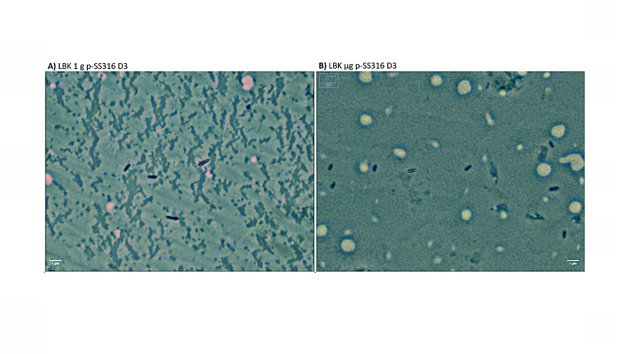
After one, two, and three days of incubation, the ISS crew terminated subsets of the experiment by fixation in paraformaldehyde, and aliquots of the supernatant were used for the planktonic cell size study presented here. The measurements were obtained post-flight through the use of phase contrast microscopy under oil immersion, a Moticam 10+ digital camera, and the FIJI image analysis program. Statistical comparisons were conducted to identify which treatments caused significant differences in cell dimensions using the Kruskal–Wallis and Dunn tests.
There were statistically significant differences as a function of material present in the culture in both LBK and mAUMg-high Pi. Along with this, the data were also grouped by gravitational condition, media, and days of incubation. Comparison of planktonic cells cultured in microgravity showed reduced cell length (from 4% to 10% depending on the material) and diameter (from 1% to 10% depending on the material) with respect to their matching Earth controls, with the caveat that the cultures may have been at different points in their growth curve at a given time.
In conclusion, smaller cells were observed on the cultures grown in microgravity, and cell size changed as a function of incubation time and the material upon which the culture grew. We describe these changes here and possible implications for human space travel in terms of crew health and potential applications.

Aliquot sample collection process, showing the BioServe’s Group Activation Pack (GAP) loaded with eight fluid processing apparatuses (FPAs) (left), an opened FPA (center) [24,26], an example coupon as used by BioServe for their studies (top right), and a 0.5 mL centrifuge conical tube as the ones sent to the Universidad del Valle de Guatemala with suspended planktonic cells (bottom right).
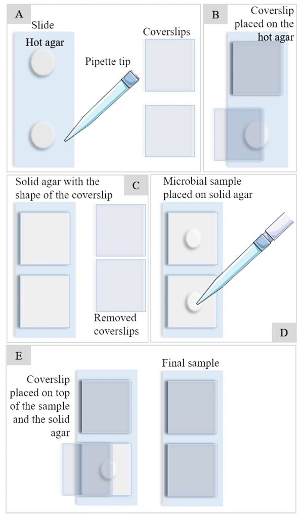
Sample preparation full process from agar bed preparation (A–C) to microbial sample pouring and fixation (D,E).
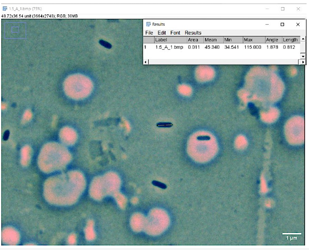
Cell measuring process. The figure shows the FIJI software and how cell length was measured. For cell diameter measurements, the same tool and process was used but the line was drawn horizontally.
Katherinne Herrera-Jordan,Pamela Pennington and Luis Zea 3
Journals Microorganisms Volume 12 Issue 2 10.3390/microorganisms12020393
https://www.mdpi.com/2076-2607/12/2/393 (open access)
Astrobiology