Tricorder Tech: Building Images Photon-by-photon To Increase The Information Content Provided By Microscopes
Editor’s note: as we develop the instruments – our tricorders – to equip Away Teams as they search for life, it is imperative that these devices be as efficient and compact as possible. Also, given the isolation of these teams, much of the initial research will likely be accomplished during a mission – thus increasing the need for the best possible data collection on field trips and back at base camp.
The world of laser scanning microscopy is quickly evolving, thanks to the advent of fast and compact detector arrays. These arrays replace the typical single-element detector of traditional confocal laser scanning microscopes, enabling new and unique capabilities. Whereas conventional detectors provide only the intensity value of the collected light, a pixelated detector also allows for recording the spatial distribution of incident light, effectively building a small image of the illuminated area for each scan point.
The extra spatial information provided by the detector arrays enable a super-resolution technique known as image scanning microscopy (ISM). ISM computationally builds a single image from a raw multidimensional dataset produced by a microscope. The final image is constructed with better signal-to-noise ratio (SNR), optical sectioning, and spatial resolution than that of a traditional confocal microscope. In detail, the lateral resolution of the ISM image can surpass Abbe’s limit up to a factor of two. These advantages, however, are achieved by exploiting only spatial information; modern fluorescence bioimaging can be further enriched by time-resolved acquisition, which enables access to structural and functional information encoded into the fluorescence dynamics (e.g., fluorescence lifetime).
Recently, researchers at the Italian Institute of Technology (IIT) in Genoa developed a compact and effective ISM microscope equipped with a single photon avalanche diode (SPAD) array detector capable of providing high-resolution structural and functional imaging in a single architecture. The research is published in the Gold Open Access journal Advanced Photonics.
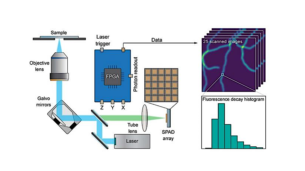
The reported SPAD array detector consists of 25 independent diodes arranged in a square grid. The small size and the asynchronous read-out enable fast detection of impinging fluorescence photons. The data acquisition scheme, based on the digital frequency domain (DFD) method, is a heterodyne sampling technique that enables the construction of the fluorescence decay histogram with a timing resolution down to 400 ps, fit for most fluorescence imaging applications. The technique is simple enough to allow for the computation of the histogram on the same field-programmable-gate-array (FPGA) board used to control the microscope and record the detected signal, simplifying the microscope architecture.
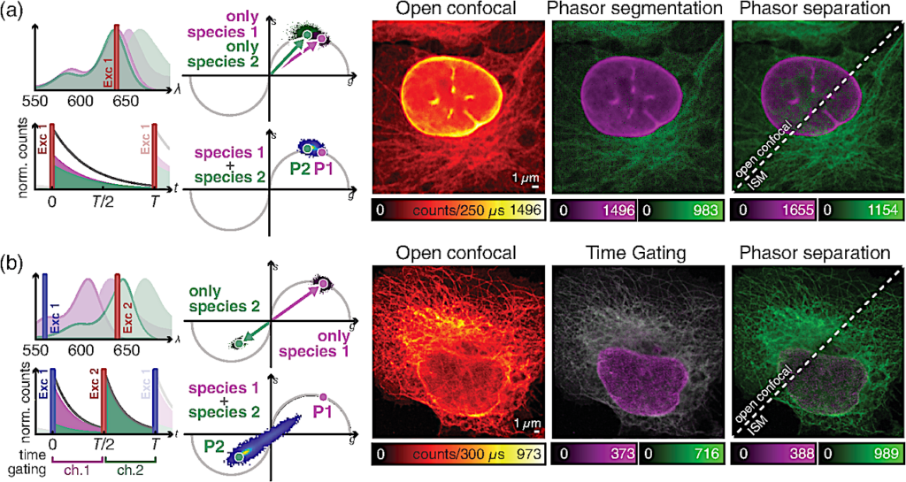
Time-resolved acquisition for multicolor ISM imaging. (a) ISM imaging of different species, reported by two dyes with similar “spectra” but different fluorescence lifetimes. (b) Pulse-interleaving ISM imaging of different species with overlapping absorption spectrum and identical lifetime. Left, schematic diagrams of (top) the absorption/emission spectra of fluorophores 1 (magenta) and 2 (green), and (bottom) of the detected signal over time. The excitation lines are also represented. Right, phasor representation of (top) two measurements obtained with only one species labeled at a time, and (bottom) of the measurement with both species labeled. Left image, raw open confocal image. Central image, open confocal result obtained by (a) segmenting the phasor space in two regions, related to the first and the second fluorophores or (b) by temporal gating of the detected signal in two temporal channels, related to the first and the second excitation events. Right image, open confocal and ISM results obtained by phasor separation.
Thanks to the unique spatial and temporal information provided by the SPAD array detector, the authors demonstrated the combination of fluorescence lifetime (FL) measurements with ISM (FLISM). In addition to the conventional ISM benefits, the improved SNR of the FLISM images enables a more robust fluorescence lifetime estimation.
The report spotlights the versatility of the microscope, by combining ISM and time-resolved measurements with stimulated emission depletion (STED) microscopy, using the separation by lifetime tuning (SPLIT) technique. The result is an image with enhanced lateral resolution and contrast, obtained without modifying the acquisition scheme. Additionally, time-resolved measurements enable multispecies imaging with a single detector for increased structural specificity.
The system can distinguish different dyes with their fluorescence lifetime values, thanks to the phasor representation of fluorescence dynamics. Even using dyes with similar lifetime values and overlapping excitation spectra, it can distinguish the different fluorophores using the pulsed-interleaving excitation technique. Indeed, by alternating excitation pulses of laser with different colours, the spectral information is effectively encoded into the temporal dimension. Thanks to the excellent time resolution of the proposed microscope, the contribution of the two fluorescent dyes can later be separated to avoid crosstalk.
According to Giuseppe Vicidomini, principal investigator at the Molecular Microscopy and Spectroscopy lab at IIT and corresponding author, “The results of this work suggest that the future of laser scanning microscopy is tightly connected to SPAD array detectors, capable of enriching the microscopy dataset with additional spatial and temporal information without the need to change the optical architecture of a confocal microscope.” The work demonstrates that SPAD array detectors combined with a tailored acquisition system make photon-resolved ISM easily accessible and usable.
For details, see the original Gold Open Access article “Compact and effective photon-resolved image scanning microscope,” Adv. Photon. 6(1) 016003 (2024) doi 10.1117/1.AP.6.1.016003.
Astrobiology, tricorder,