Tricorder Tech: Quantitative Measurement Of Microbial Growth Rate With Raman Microspectroscopy
Rates of microbial activity and growth are fundamental to understanding environmental geochemistry and ecology.
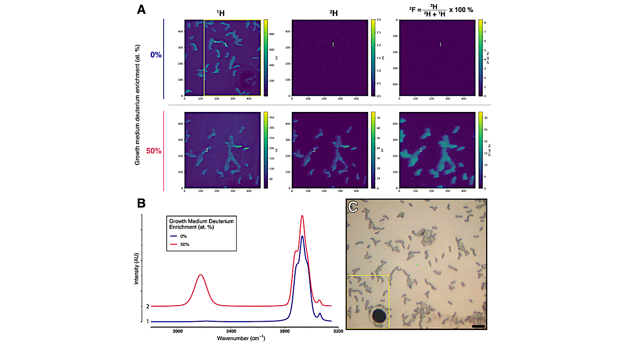
However, measuring the heterogeneity of microbial activity at the single-cell level, especially within complex populations and environmental matrices, remains a forefront challenge. Stable Isotope Probing (SIP) is a standard method for assessing microbial activity and involves measuring the incorporation of an isotopically labeled compound into microbial biomass.
Here, we assess the utility of Raman microspectroscopy as a SIP technique, specifically focusing on the measurement of deuterium (2H), a tracer of microbial biomass production. We generate calibrations of microbial biomass 2H values and find that Raman microspectroscopy reliably quantifies 2H incorporation ranging between 0 and 40 at. %.
Applying the results of this calibration to a SIP model, we explicitly parameterize the factors controlling microbial growth quantification, demonstrating how Raman-SIP can measure the growth of microorganisms with doubling times ranging from hours to years.
Furthermore, we correlatively compare our Raman-derived measurements with those of nanoscale secondary ion mass spectrometry (nanoSIMS) to compare the relative strengths of nanoSIMS- and Raman-based SIP approaches.
We find that Raman microspectroscopy is a robust, accessible methodology that can readily differentiate and quantify the growth of individual microbial cells in complex samples.
Quantitative measurement of microbial growth rate with Raman microspectroscopy, biorxiv.org
Astrobiology