Tricorder Tech: Android-based Application For Photoacoustic Tomography Image Reconstruction
Editor’s note: Photoacoustic tomography allows unique capabilities to look deep inside an organism. Up until now such a capability required the use of a desktop computer to process the data generated by observations. This new capability utilizes a smartphone format and is portable. While the sensor set-up itself is still not especially portable, the computing power to process the data it collect fits in your pocket. One more step toward the sort of capability you might want to have in a Tricorder during an Away Team expedition. We’ll be keeping an eye on this technology.
Photoacoustic tomography (PAT) is a hybrid imaging technique that combines optical illumination with ultrasound detection for high-resolution imaging of deep tissues. Utilizing the photoacoustic (PA) effect, PAT provides a distinct advantage with scalable resolution, higher imaging depth, and high contrast imaging. It uses nanosecond laser pulses to illuminate the tissue of interest, enabling their chromophores to absorb the incident laser energy. This results in a local temperature rise and generates pressure waves that propagate to the tissue boundary as ultrasound waves. These PA waves are then acquired with the help of an ultrasound transducer and converted into internal absorption maps using reconstruction algorithms.
This process of generating initial pressure maps can be carried out with several different image reconstruction algorithms, including the simple delay-and-sum (DAS) beamformer. This algorithm back-projects the acquired signals from various tissue locations, which are then added at every pixel in the reconstructed image. However, this makes the DAS beamformer computationally expensive and time-consuming, and results in artifacts, i.e., anomalies, in the reconstructed images. Despite these drawbacks, its simplicity and ease of implementation make it a popular choice for PAT reconstruction.
Typically, implementing such reconstruction algorithms requires a workstation, desktop, or laptop with extensive computational resources. But the computational power of mobile phones has been growing in recent years. Although mobile phones have been proposed for various microscopy modalities including ultrasound imaging, their utility for photoacoustic imaging such as PAT image reconstruction has not been explored.
Capitalizing on the advanced processing ability of mobile phones, researchers from Singapore and the United States have now developed an Android-based application for PAT image reconstruction. The study was led by Manojit Pramanik, Northrop Grumman Associate Professor in the Department of Electrical and Computer Engineering at Iowa State University, and published in the Journal of Biomedical Optics (JBO).

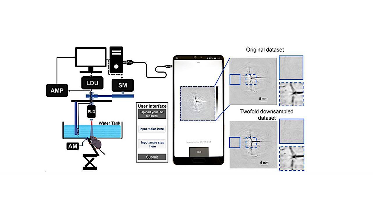
The developed application utilizes a single-element ultrasound transducer (SUT)-based DAS beamformer algorithm for image reconstruction on Kivy—a cross-platform Python 3.9.5 framework.
The researchers verified its performance on different mobile phones by using simulated and experimental PAT datasets. While the simulated datasets consisted of point targets, triangular shape, and rat’s brain vessel shape, experimental datasets comprised of a point source phantom, a triangular shape phantom and blood vessels in the brain of live rats.
“The developed application can successfully reconstruct the PAT data into high-quality PAT images with signal-to-noise ratio values above 30 decibels,” comments Pramanik.
Interestingly, the algorithm’s computational time on a Huawei P20 mobile phone was comparable to that on a laptop for small datasets. Furthermore, two-fold downsampling of the original dataset reduced the computational time while maintaining the image quality, thus allowing image reconstruction with both speed and quality. In contrast, three-fold downsampling visibly degraded the PAT images.
Moreover, the researchers found that with the Samsung Galaxy S21+’s advanced processor, PAT reconstruction could be achieved in only 2.4 seconds. “This is a considerably reduced running time for image reconstruction and highlights the efficiency of the mobile phone application,” notes Pramanik.
JBO Editor-in-Chief Brian Pogue, Chair of Medical Physics at University of Wisconsin–Madison, remarks, “This first-of-its-kind application provides an opportunity for PAT image reconstruction on inexpensive, portable, and widely available mobile phones. Going ahead, the application can make PAT systems more adaptable and extendable to other fields of biomedical imaging, facilitating point-of-care diagnosis.” He adds, “The code for this Android-based application has been made freely available on GitHub, making this a major service to the biomedical imaging community.”
Read the Gold Open Access article by Hui et al., “Android mobile-platform-based image reconstruction for photoacoustic tomography,” J. Biomed. Opt. 28(4), 046009 (2023), doi 10.1117/1.JBO.28.4.046009.
Astrobiology