Tricorder Tech: 3D Printed Compact Imaging Platform Validated for Space Research with C. elegans
In this newly published article, Dr. Siva Vanapalli of Texas Tech University reports on the testing and performance of a 3D printed compact imaging platform (CIP) that is integrated with a smart-device camera for the whole-organism phenotyping of the model nematode worm C. elegans.
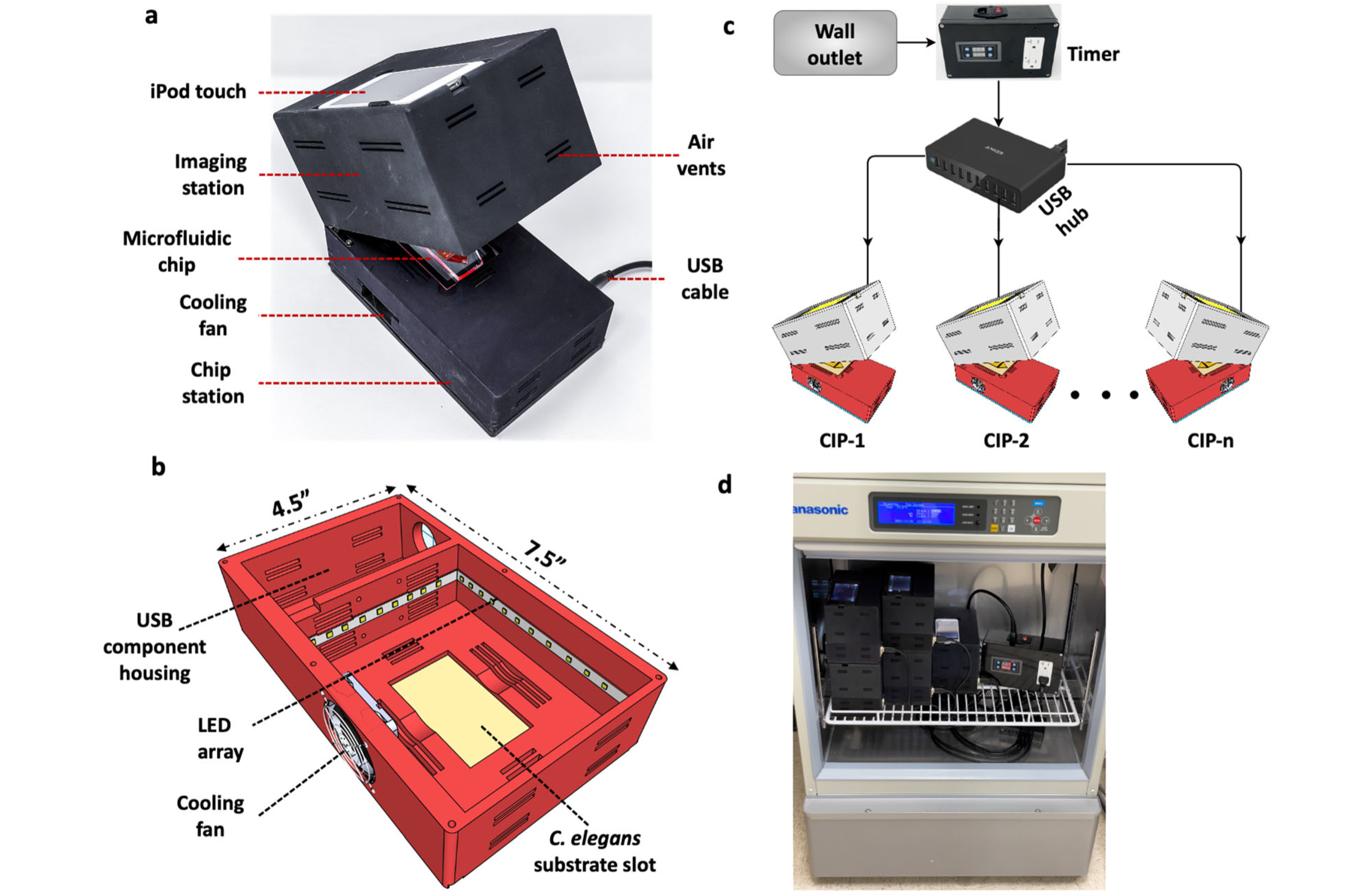
The CIP has no external optical elements and does not require mechanical focusing, simplifying the optical configuration. The small footprint of the system powered with a standard USB provides capabilities ranging from plug-and-play, to parallel operation, and to housing it in incubators for temperature control.
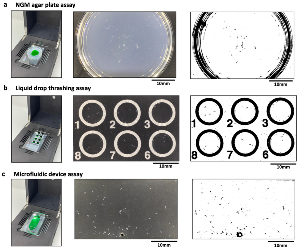
Compatibility of the CIP with various C. elegans substates. (a) Imaging of animals crawling on a 3 cm agar plate (left). The actual (middle) and processed (right) image are also shown with n = 24 animals. The green dye region indicates the bacterial lawn. (b) Imaging of animals thrashing in liquid drops (left). The actual (middle) and processed (right) image are also shown with n = 3 animals per drop. The field of view allows imaging of 6 wells. (c) Imaging of animals crawling in a microfluidic pillar arena (left). The actual (middle) and processed (right) image are also shown with n = 51 animals in the arena. The images were processed using Image J V 1.53 (NIH, Maryland, USA), wrMTrck plugin.
Dr. Vanapalli and his team demonstrates the compatibility of the CIP with different C. elegans substrates, including agar plates, liquid droplets on glass slides and microfluidic chips. The system has been validated with behavioral and thrashing assays and shows that the phenotypic readouts are in good agreement with the literature data. A pilot study was conducted using mutant strains of C. elegans, which shows that the phenotypic data collected from the CIP successfully distinguishes these mutants. Results from this study demonstrate the compactness, portability and ease-of-use makes the CIP desirable for research and educational outreach applications on Earth and in space.
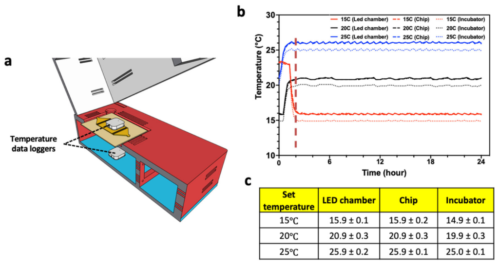
Controlling temperature of CIP. (a) Temperature was measured at the top of the microfluidic chip and in the LED chamber. (b) Temperature profile collected over 24 h at set incubator temperatures of 15 °C, 20 °C, and 25 °C. The temperature data of LED chamber and the microfluidic chip are nearly identical. (c) Table showing mean temperature and standard deviation from all the three collection points.
The model organism Caenorhabditis elegans is used in a variety of applications ranging from fundamental biological studies, to drug screening, to disease modeling, and to space-biology investigations. These applications rely on conducting whole-organism phenotypic assays involving animal behavior and locomotion. The simplicity and versatility offered by the compact imaging platform tested in this study makes it amenable to future C. elegans investigations on the International Space Station, where science experiments are constrained by system size, payload weight and crew time. The article is available online here: A Compact Imaging Platform for Conducting C. elegans Phenotypic Assays on Earth and in Spaceflight, Life (open access)
This study was funded by Space Biology grant, “Determining Muscle Strength in Space-Flown Caenorhabditis elegans” to Dr. Siva Vanapalli of Texas Tech University. Dr. Vanapalli is a professor in the Chemical Engineering Department where he studies microfluidics, fluid mechanics of drops, polymer solutions and cells, tumor cell mechanics and cancer diagnostics, devices for drug screening and for C. elegans biology.
Astrobiology, Tricorder, space biology