Tricorder Tech: FLASHlight MRI In Real Time—A Step Towards Star Trek Medicine

Editor’s note: If you search for a definition for tricorder you’ll find things like this from Wikipedia: “A tricorder is a fictional handheld sensor that exists in the Star Trek universe. The tricorder is a multifunctional hand-held device that can perform environmental scans, data recording, and data analysis; hence the word “tricorder” to refer to the three functions of sensing, recording, and computing.”
You’ll also see the term openly used for contemporary medical devices that are used to do quick tests of someone’s health or some mineral’s composition. In Star Trek a physician usually had a smaller device that was connected to a larger tricorder to scan humans for their health status.
In an era of ubiquitous smartphones we’re now expecting more and more functionality with every new model that is released. As we move closer to sending humans to other worlds to search for life, such a device that falls under the general “tricorder” classification would certainly come in handy. These Astrobiology Away Team sorties will find it very useful to look inside the things that they observe during their traverses – living and not living.
This work describes a dynamic magnetic resonance imaging (MRI) technique for local scanning of the human body with use of a handheld receive coil or coil array. Real-time MRI is based on highly undersampled radial gradient-echo sequences with joint reconstructions of serial images and coil sensitivity maps by regularized nonlinear inversion (NLINV).
For this proof-of-concept study, a fixed slice position and field-of-view (FOV) were predefined from the operating console, while a local receive coil (array) is moved across the body—for the sake of simplicity by the subject itself.
Experimental realizations with a conventional 3 T magnet comprise dynamic anatomic imaging of the head, thorax and abdomen of healthy volunteers. Typically, the image resolution was 0.75 to 1.5 mm with 3 to 6 mm section thickness and acquisition times of 33 to 100 ms per frame. However, spatiotemporal resolutions and contrasts are highly variable and may be adjusted to clinical needs.
In summary, the proposed FLASHlight MRI method provides a robust acquisition and reconstruction basis for future diagnostic strategies that mimic the usage of ultrasound. Necessary extensions for this vision require remote control of all sequence parameters by a person at the scanner as well as the design of more flexible gradients and magnets.
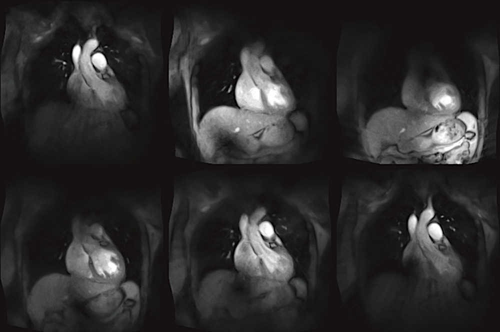
Selected T1-weighted images of a 60 s FLASHlight MRI acquisition (11 cm loop coil) covering the thorax of a healthy subject at 33.3 ms temporal resolution. For further details see Table 1. MRI, magnetic resonance imaging. — Quant Imaging Med Surg
FLASHlight MRI in real time—a step towards Star Trek medicine, Quant Imaging Med Surg. 2022 Oct 8;13(1):489–495. doi: 10.21037/qims-22-648 via PubMed (open access) – with videos
See: Tricorder Tech: That Star Trek Medical Scanner Is Getting Much Closer
Astrobiology