Microfossils Shed Light On The Long Fossil Record Of Euglenoids
Hiding in the shadows, euglenoids are a fascinating group of single-celled protists that are neither plant nor animal.
Plants photosynthesize, and animals eat. Euglenoids do both. Spiraling along the murky bottoms of shallow fresh-water ponds with their long flagella, they eat organic goop, while also using their chloroplasts to convert CO2 and water with light into sugars. Because of this in-between status, euglenoids have been placed close to the very base of the eukaryotic branch on the tree-of-life that includes all plants, fungi, and animals. However, while euglenoids likely evolved more than 1 billion years ago, they appeared to have left only a very scant fossil record.
In a new study published in the journal Review of Palaeobotany and Palynology, a team of Dutch, American, British, German, and Australian scientists shed new light on a group of “problematic” microfossils that have remained a mystery for nearly a century. By comparing microscopic fossil cysts in 200-million-year-old pond sediments from cores drilled in Germany and the Netherlands to much older Paleozoic, and much younger remains in Holocene lakes in Greece, and finally to living protists in a pond in Australia, the researchers establish a 400-million-year evolutionary history of the euglenoids.
What’s in a name?
In 2012, Bas van de Schootbrugge, then at the Goethe University in Frankfurt am Main, and Paul Strother from Boston College, while working on a variety of problematic microfossils in sediments from around the Triassic-Jurassic boundary, realized that the circular striated cysts they were seeing, could be in fact euglenoid cysts. “We had this amazing drill core material at our disposal that contained many unidentified microfossils, including some of the oldest butterfly remains that we published on in 2018”, said Bas van de Schootbrugge, now at Utrecht University. Paul Strother continued: “Some of the microfossils we encountered showed a canny similarity to cysts of Euglena, a modern representative that had been described by Slovakian colleagues. The problem was, there was only one publication in the world making this claim”.
Even more unsettling: after an extensive literature review, van de Schootbrugge and Strother realized that the same type of microfossil had been given many different names. Scientists working on Quaternary and Holocene time slices used Concentricystes, referring to a possible algal cyst with concentric ribs. But Mesozoic workers used Pseudoschizaea, originally thinking it could have been a fern spore. Even older fossils from the Permian, Devonian, Silurian and Ordovician were known as Circulisporites and Chomotriletes.
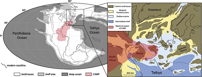
Reconstructed palaeographic map of the Triassic-Jurassic boundary interval. The map insert shows the location of the four sampled cores in Europe: (1) Schandelah-1 core (Germany), (2) Winterswijk core (Netherlands), (3) Boust core, (France), (4) Mingolsheim core (Germany). Map is modified from van de Schootbrugge et al. (2020) and Bos et al. (2023).
Transmission electron microscopy
After the authors had disentangled the taxonomic confusion, compiling in the process nearly 500 literature sources related to any of the four taxa, more advanced microscope techniques were needed to establish the ultrastructure of the cysts with the help of transmission electron microscopy (TEM). This required picking of single specimens, embedding, and micro-tome slicing by University of Wisconsin-Eau-Claire co-author Wilson Taylor. Because the specimens in the Triassic-Jurassic cores were mostly damaged, the team turned to palynologist Andreas Koutsodendris at Heidelberg University (Germany), who had access to Holocene and Pliocene core samples containing abundant well-preserved specimens.
Andreas Koutsodendris said: “I am encountering these cysts regularly in cores drilled in lakes, for example in Lake Vouliagmeni in Greece that we studied here, but their biological affinity has never been cleared. In fact, the cysts are commonly figured in publications by colleagues, but no one was able to really put a finger on it.” Wilson Taylor continued: “We were much surprised by the ultrastructure of the cysts. The structure of the wall does not resemble anything that is known. The ribs are not ornaments, like in pollen and spores, but part of the wall structure”, said Wilson Taylor. “The layered structure of the walls is also clearly different from many other fresh-water green algae”, Taylor continued.
Nagging uncertainty
While the TEM analysis initially added more mystery, the results did align with a study published in 2021 by another group of colleagues that looked at the ultrastructure of Pseudoschizaea. At least it was possible to show that Holocene and Pliocene Concentricystes and Jurassic Pseudoschizaea are in fact the same. But there remained one nagging uncertainty and that was the lack of any cysts produced by living euglenoids. Wilson Taylor: “We did contact several biologists working on living euglenoids, but no one had been able to make euglenoids encyst in a lab setting, allowing for extraction and TEM analyses of the cysts”.
Microscopic life down under
Enter Fabian Weston. By chance, Strother and van de Schootbrugge stumbled across superb video material posted on YouTube by microscopy enthusiast Fabian Weston from Sydney, Australia. In 2020 Fabian Weston had put a drop of water sampled from a nearby pond in New South Wales on a microscope slide, and using his advanced set-up at The Protist Lab filmed Euglena as it gracefully moved in and out of focus. For reasons that remain poorly understood but could be related to the drying out of the water under the cover slip, Euglena is then seen to ball-up and form a thick wall with ribs that is akin to the cysts found throughout the fossil record. “Unwittingly, Fabian provided a key piece of evidence. He is probably the only person on the planet to have witnessed Euglena encyst under a microscope”, Strother said.
Significance and next steps
Based on all the available pieces of the puzzle, the authors thus link euglenoids from a pond in Australia to fossil cysts that are more than 400 million years old, establishing a deep time record of the euglenoids. “This opens the door for recognizing even older examples, for example from Precambrian records that go back to the very root of the eukaryotic tree of life”, Strother said. “Now that we know which organisms produced those cysts, we can also use them for paleo-environmental interpretations. Their abundance around two of the largest mass-extinction events of the past 600 million years is a tell-tale sign of some major upheavals on the continents related to increased precipitation under extreme greenhouse climate conditions.”
Van de Schootbrugge concluded: “Perhaps related to their capabilities to encyst, these organisms have endured and survived every major extinction on the planet. Unlike the behemoths that were done in by volcanoes and asteroids, these tiny creatures have weathered it all.” Extending their research, the team intends to travel to Australia in the near future to scour for preserved Euglena cysts in pond and lake sediments in New South Wales.
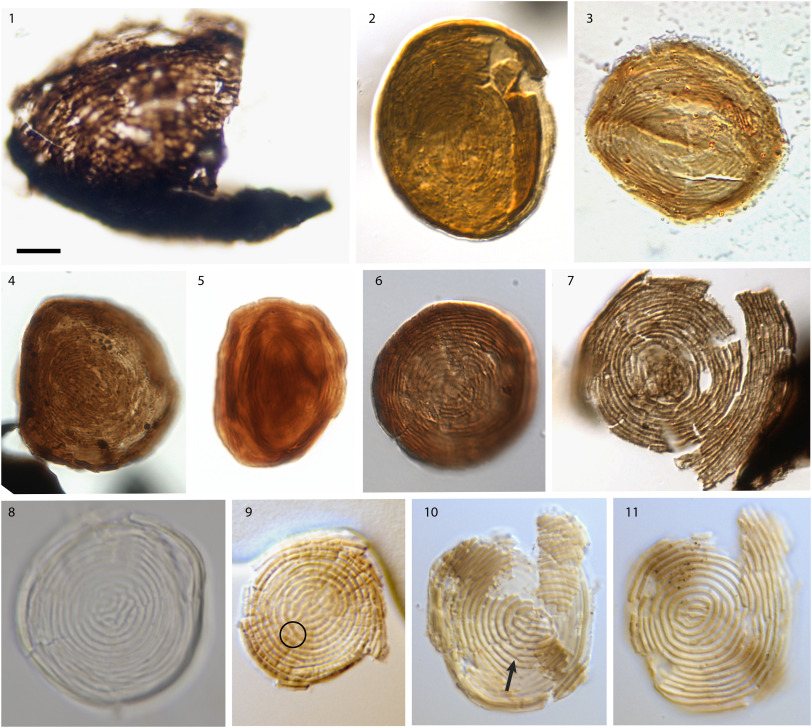
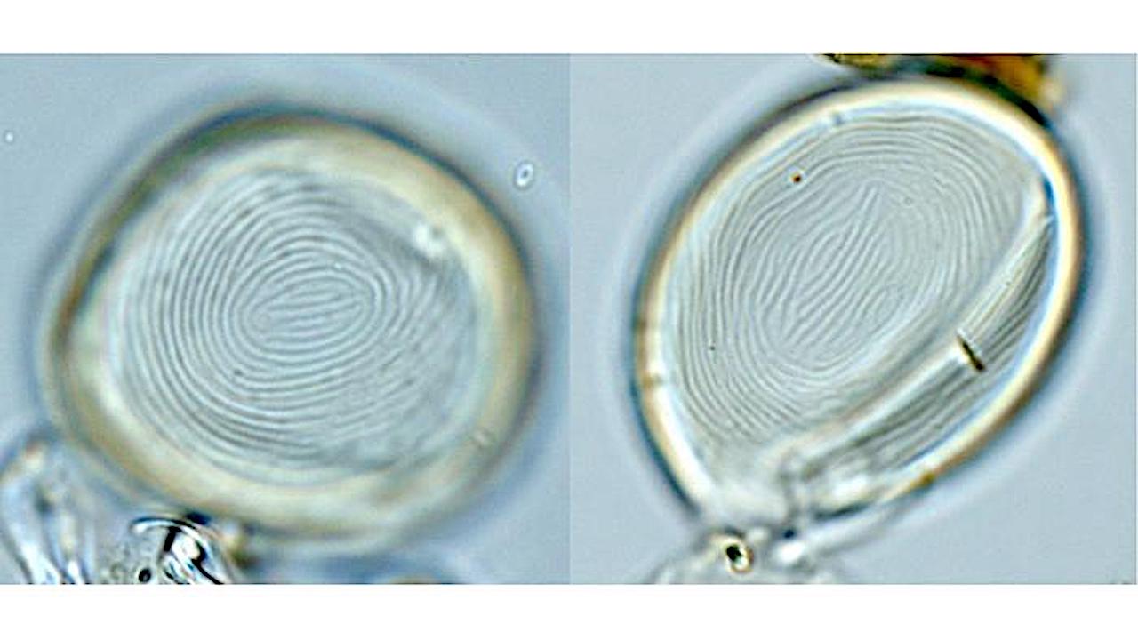
Stratigraphical examples of Chomotriletes s.l. 1. Chomotriletes sp. Illion Shale (Utica, New York, USA); sample S14–2, Silurian (Wenlock). 2. Pseudoschizaea sp., Winterswijk core (Netherlands) WINT15–02; sample 15.5 mbs, upper Rhaetian (Triassic) 3. Pseudoschizaea sp. Boust core (N France); sample BOUST_44_58 from the Argiles de Levallois, upper Rhaetian (Triassic). 4. Pseudoschizaea sp. Schandelah-1 core (N Germany); sample SCH_338_00 from the Contorta Schichten, middle Rhaetian (Triassic). 5 Pseudoschizaea sp. Schandelah-1 core (N Germany); sample SCH_338_00 from the Contorta Schichten, middle Rhaetian (Triassic). 6. Pseudoschizaea sp. Schandelah-1 core (N Germany); sample SCH_335_50 from the Contorta Schichten, middle Rhaetian (Triassic). 7. Pseudoschizaea sp. Mingolsheim core (S Germany); sample MING41A from the Triletes Beds, uppermost Rhaetian (Triassic). 8. Concentricystes sp. from Lake Vouliagmeni, Greece. (Holocene). 9. Concentricystes sp. IODP core U1478, Mozambique Channel (Pliocene), showing truncated costae (circle). 10. Concentricystes sp. IODP core U1478, Mozambique Channel (Pliocene), showing costae branching (arrow). 11. Concentricystes sp. IODP core U1478, Mozambique Channel (Pliocene).
Recognition of an extended record of euglenoid cysts: Implications for the end-Triassic mass extinction, Review of Palaeobotany and Palynology (open access)
Astrobiology,