An Update On Eukaryotic Viruses Revived From Ancient Permafrost
One quarter of the Northern hemisphere is underlain by permanently frozen ground, referred to as permafrost.
Due to climate warming, irreversibly thawing permafrost is releasing organic matter frozen for up to a million years, most of which decomposes into carbon dioxide and methane, further enhancing the greenhouse effect.
Part of this organic matter also consists of revived cellular microbes (prokaryotes, unicellular eukaryotes) as well as viruses that remained dormant since prehistorical times. While the literature abounds on descriptions of the rich and diverse prokaryotic microbiomes found in permafrost, no additional report about “live” viruses have been published since the two original studies describing pithovirus (in 2014) and mollivirus (in 2015). This wrongly suggests that such occurrences are rare and that “zombie viruses” are not a public health threat.
To restore an appreciation closer to reality, we report the preliminary characterizations of 13 new viruses isolated from 7 different ancient Siberian permafrost samples, 1 from the Lena river and 1 from Kamchatka cryosol. As expected from the host specificity imposed by our protocol, these viruses belong to 5 different clades infecting Acanthamoeba spp. but not previously revived from permafrost: pandoravirus, cedratvirus, megavirus, and pacmanvirus, in addition to a new pithovirus strain.
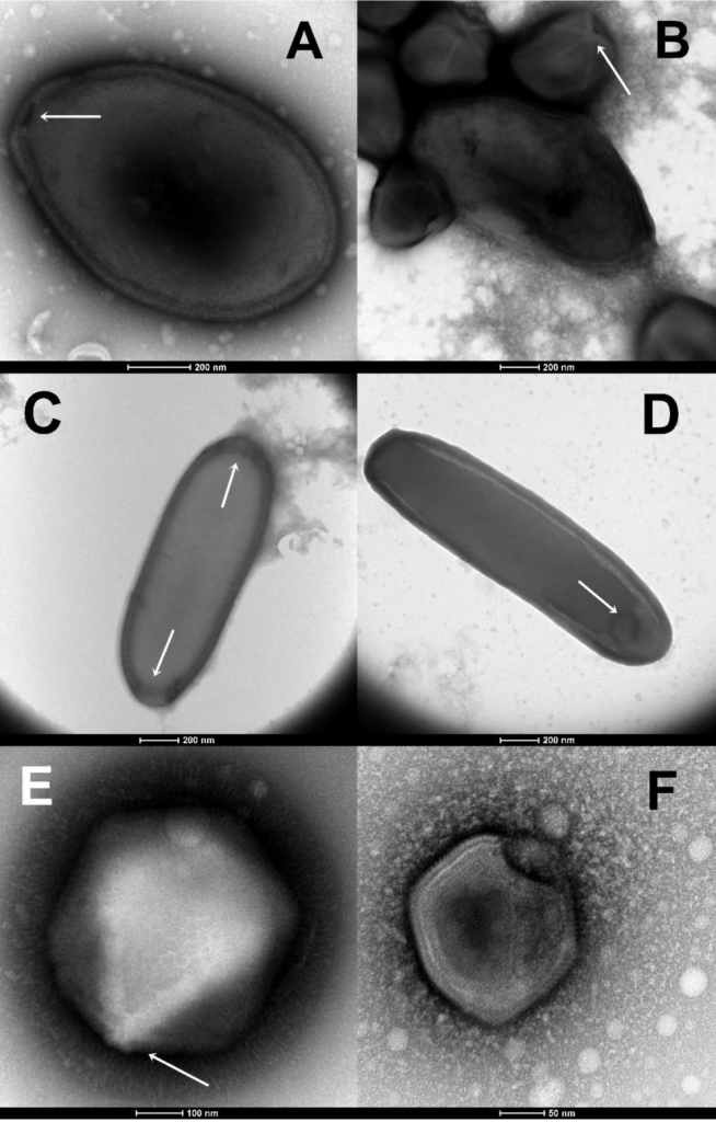
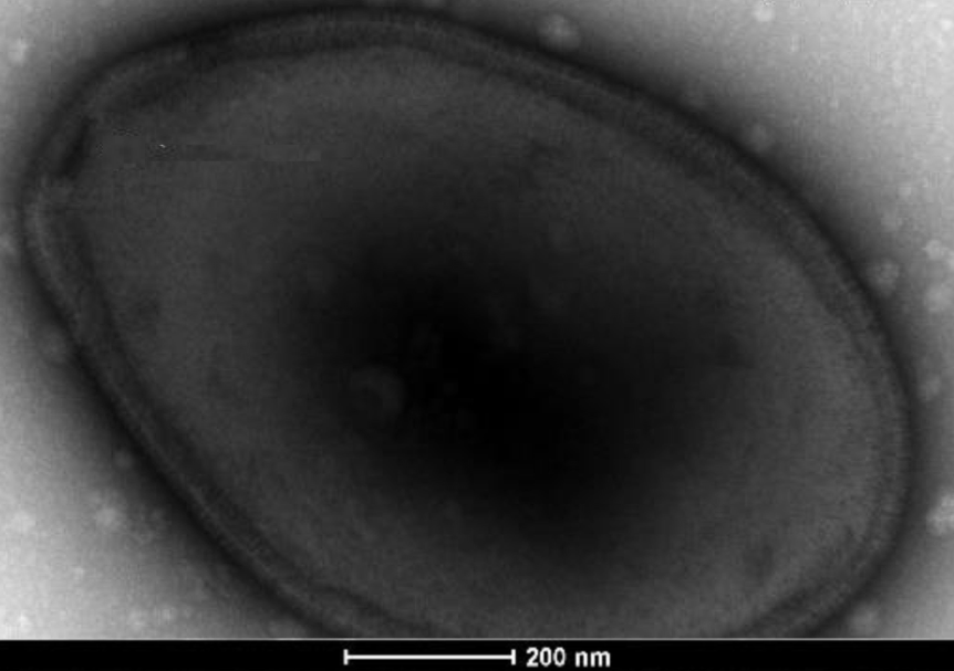
Morphological features guiding the early identification of newly isolated viruses (Negative staining, Trans- 203 mission Electron Microscopy). (A) The large ovoid particle (1,000 nm in length) of Pandoraviruses with its characteris- 204 tic apex ostiole (white arrowhead). (B) A mixture of Pandoravirus particles and Megavirus icosahedral particles ex- 205 hibiting a “stargate” (white starfish-like structure crowning a vertex, white arrowhead). (C) The elongated particle of 206 a Cedratvirus (1,500 nm in length) exhibits two apex cork-like structures (white arrowheads). (D) The elongated parti- 207 cle of a Pithovirus (1,900 nm in length) exhibits a single apex cork-like structure (white arrowhead). (E) The large (770 208 nm in diameter) “hairy” icosahedral particle of a Megavirus, with its prominent “stargate” (whaite arrowhead). (F) 209 The smaller icosahedral particle (200 nm in diameter) typical of Asfarviruses/Pacmanviruses. — biorxiv
Jean-Marie Alempic, Audrey Lartigue, Artemiy E Goncharov, Guido Grosse, Jens Strauss, Alexey N. Tikhonov, Alexander N. Fedorov, Olivier Poirot, Matthieu Legendre, Sébastien Santini, Chantal Abergel, Jean-Michel Claverie
doi: https://doi.org/10.1101/2022.11.10.515937
https://www.biorxiv.org/content/10.1101/2022.11.10.515937v1
Full paper
Astrobiology