Origin Of Photosynthesis In A Sun-loving Bacteria

Every day, enough sunlight hits the Earth to power the planet many times over — if only we could more efficiently capture all the energy.
With today’s solar panels limited by their efficiency (currently, more than 80 percent of available solar energy is lost as heat), scientists have been looking into nature as inspiration to better understanding the way photosynthetic plants and bacteria capture sunlight.
“Nature’s invention of photosynthesis is the single most important energy conversion process driving the biosphere, and photosynthesis forever changed the Earth’s atmosphere,” said Raimund Fromme, an associate research professor at the ASU Biodesign Institute’s Center for Applied Structural Biology and in the School of Molecular Sciences.
More than 3 billion years ago our planet had an atmosphere without oxygen. At this time, nature figured out a way to capture the sunlight and convert it food to take advantage of this everlasting energy source.
Now, a research group led by Fromme has gained important new insights by resolving with near-atomic clarity, the very first core membrane protein structure in the simplest known photosynthetic bacterium, called Heliobacterium modesticaldum (Helios was the Greek sun god).
By solving the heart of photosynthesis in this sun-loving, soil-dwelling bacterium, Fromme’s research team has gained a fundamental new understanding of the early evolution of photosynthesis, and how this vital process differs between plants systems.
Their discovery provides scientists with a brand new template for laying the groundwork for organic-based solar panel design, known as “artificial leaves” for solar energy, or possible renewable biofuel applications.
The findings appear in today’s issue of Science.

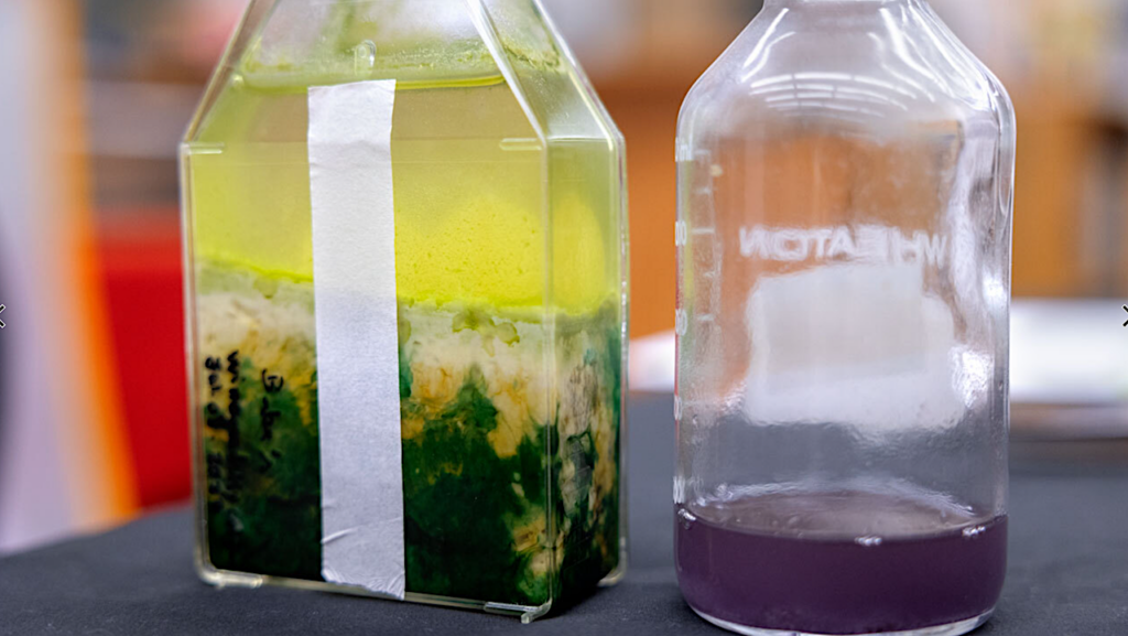
Fromme’s research project was started seven years ago as postdoctoral researcher Iosifina Sarrou first improved the preparation of the heliobacterial reaction center. After many initial trials of crystallization, an X-ray diffracting crystal charge was found. “This is the moment a crystallographer is waiting for,” said Raimund Fromme, explaining the years it can take to grow the perfect protein crystal suitable for X-ray studies (shown on the left). The figure on the right is the diffraction pattern after the crystals were exposed to X-rays. Scientists can understand the 3-D structure of a protein from these patterns. CREDIT ASU
Nature knows best
ASU has long been a long-standing research leader in photosynthesis, going back to its first decade as a research university in the 1970s. It was a natural fit for scientists drawn to the unique beauty of the Sonoran Desert that, with its 300-plus days of annual sunshine, is simply the best spot in the nation to capture solar energy.
Life’s solar panels, which scientists call photosystems, are used by plants, algae and photosynthetic bacteria as an incredibly efficient system for capturing almost every available photon of light to grow and thrive, filling almost every nook and cranny on earth.
Fromme is part of a large structural biology research group at ASU, who are steadily gaining a better understanding by taking pictures of the key proteins that work within the photosynthesis reaction centers to help turn light into energy.
“To truly and fully understand photosynthesis, one has to follow the process of converting light into chemical energy,” said Fromme. “This is one of the fastest chemical reactions ever studied, which is part of what makes it so hard to study and understand.”
The timescales of photosynthesis turn a bolt of lightning into a snail-like pace by comparison. Photosynthesis reactions occur at the scale of picoseconds, which is one-trillionth of a second. A picosecond is to one second as one second is to 37,000 years.
But the ASU structural biologists are using ever more powerful x-ray technology to one day catch up to the light by capturing freeze-frame images of crystallized proteins throughout the whole process.

Using X-ray light at the Advanced Light Source in Berkeley, CA, and a beamline at Advanced Photon Source at Argonne National Lab, IL., Fromme’s group has now visualized the heliobacteria reaction centers for the very first time at near-atomic, 2.2-angstrom resolution (an angstrom is the width of a hydrogen atom). They found an almost perfect symmetry in the heliobacter RC (shown by the mirror image pattern of the main protein motifs, indicated by cylinders (top image) and a close up of the exact placement of individual reaction center atoms (bottom). CREDIT ASU
Lightning in a bottle
In an effort to study photosynthesis, Fromme explored photosynthesis in its simplest form, in heliobacteria, which were first found in muddy soils near hot springs.
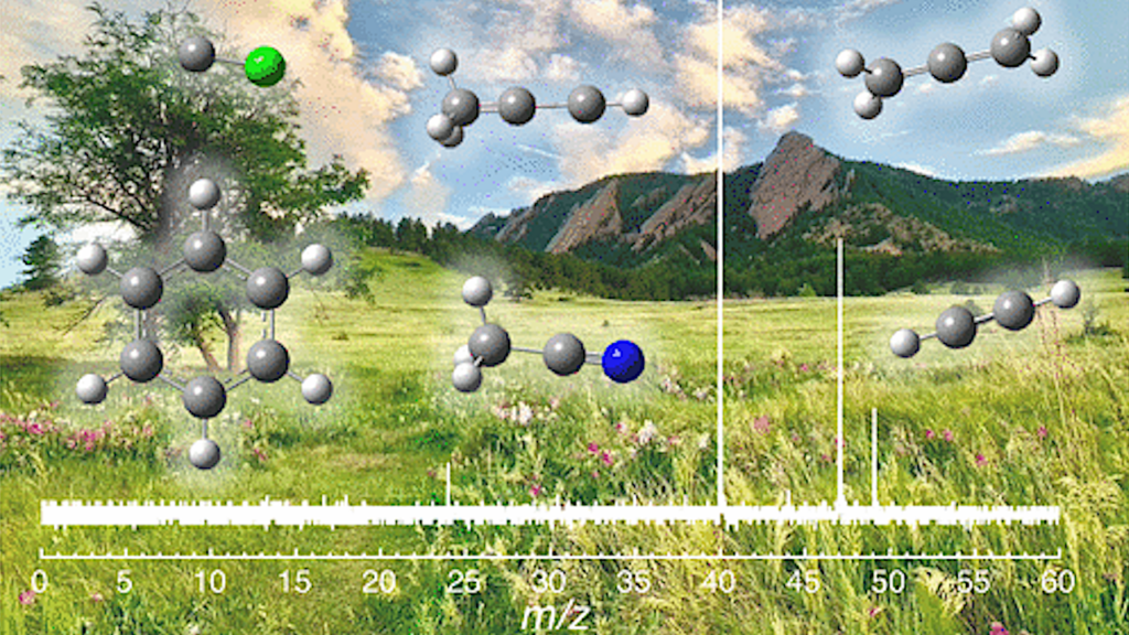
Single-celled heliobacteria are simpler, yet fundamentally different than plants. For instance, during photosynthesis, instead of using water like plants, heliobacteria use hydrogen sulfide. They grow without oxygen, and after photosynthesis, give off a rotten-egg smelling sulfur gas in place of oxygen.
Heliobacteria have used their unique place to successfully carve out their own ecological niche because they use a near-infrared wavelength of light for photosynthesis, which is perfect for low light conditions found in places like Iceland or muddy water rice paddies. Plants simply can’t compete.
Scientists have wanted to understand how heliobacteria accomplish this.
Reaction to action
At the heart of photosynthesis is a reaction center; it’s an elaborate complex of pigments and proteins that turn light into electrons to power the cell.
Chlorophyll is the pigment that makes plants green. In plants, chlorophyll captures the sun’s energy and uses it to make sugars out of carbon dioxide from the air and water.
Oxygenic photosynthesis in higher plants, green algae and cyanobacteria make use of Photosystem I (PSI), which is a Type I RC, and Photosystem II (PSII), which is a Type II RC. These work together to extract electrons from water to ferredoxin and finally reduce an energy carrier NADP+ to NADPH.
In contrast, anoxygenic phototrophic bacteria, such as Heliobacterium modesticaldum, use a single RC to drive a cyclic electron transfer (ET) pathway that creates a proton-motive force across the membrane, which is used to drive energy production and metabolism by ATP synthesis.
The reaction centers enclose these participants like a cage to efficiently capture all the available energy and photons of light by bringing all the elements together in the same vicinity.
Reaction centers (RC) come in two main flavors of cofactors: iron (Type I) or quinone (Type II).
Heliobacteria have the simplest known reaction center and use unique chlorophylls.
The discovery of heliobacteria led to the identification of unique characteristics for its RC (in the 1990s, former ASU chemistry chair Robert Blankenship first led a group to help sequence and characterize heliobacteria RCs).
The heliobacteria RC has been proposed to be the closest thing alive to the earliest common ancestor of all photosynthetic reaction centers, when, around 3 billion years ago, the early Earth contained sulfur rich seas and little oxygen.
But successfully purifying an RC protein and growing crystals needed for X-ray experiments can be a lengthy, difficult process.
In particular, Fromme’s research project was started seven years ago as postdoctoral researcher Iosifina Sarrou first improved the preparation of the heliobacterial reaction center. After many initial trials of crystallization,
an X-ray diffracting crystal charge was found.
“This is the moment a crystallographer is waiting for,” said Fromme, explaining the years it can take to grow the perfect protein crystal suitable for X-ray studies.
Two to tango
Short after these encouraging results, Christopher Gisriel joined the team and improved the diffraction quality to the final quality of 2.2 Angstroms.
Still, the research team could not solve a crystallographic structure.
This hiatus took two years until August 2016. Then, finally, a breakthrough came.
At this point “a thrilling discovery on unchartered territory began, as each new chlorophyll was cheered,” Fromme remembered, and “proved everyone’s initial prediction on the heliobacteria’s RC was wrong.”
Using X-ray light at the Advanced Light Source in Berkeley, CA, and a beamline at Advanced Photon Source at Argonne National Lab, IL., Fromme’s group has now visualized the heliobacteria RCs for the very first time at near-atomic, 2.2-angstrom resolution (an angstrom is the width of a hydrogen atom).
They found an almost perfect symmetry in the heliobacter RC.
First, the amino acid composition of a pair of proteins was identical, called a homodimer.
This was the very first time that a RC was found to contain just a single pair of protein homodimers to drive photosynthesis.
Finally, they mapped about 60 chlorophylls onto the RC protein complex, which was finally a way higher number than his colleague John Golbeck from Pennstate University who was part of the study predicted.
The core polypeptide dimer and two small subunits coordinate 54 (bacterio)chlorophylls and 2 carotenoids that capture and transfer energy to the core at the reaction center, which performs charge separation, stabilization and electron transfer it consists of 6 (bacterio)chlorophylls and an iron-sulfur cluster; unlike other reaction centers, it lacks a bound quinone.
Thus, the structure supports the hypothesis that electron transport in the HbRC does not require an intermediate cofactor.
“High-resolution structures have been obtained from multiple heterodimeric (more than one protein) RCs (Purple bacteria RC, PSI, and PSII), but no homodimeric RC structure have been solved until now,” said Fromme.
The ancient origins of photosynthesis
Furthermore, with the explosion of DNA sequencing technology, and with the potential ability to understand all of the genes and proteins across life, they also traced the evolution of the photosynthesis RCs.
Could this reaction center have spawned all others, leading to greater complexity over the eons?
In evolutionary terms, this means that the heliobacteria RC may have first come from a single gene.
“This structure preserves characteristics of the ancestral reaction center, providing insight into the evolution of photosynthesis,” explains Fromme’s colleague Kevin Redding. “From the new structures we have, it would certainly make sense for a compelling case.”
Then, the gene may have been duplicated to increase the evolutionary complexity.
“A homodimeric RC almost certainly preceded heterodimeric RCs in evolution,” said Fromme. Duplication of the core RC subunit gene followed by divergence of the two genes would allow for the conversion of a homodimeric to a heterodimeric RC. This likely occurred on at least three separate occasions, leading to the creation of all the different and more complex reactions center found in other photosynthetic bacteria and plants.”
The sun will rise again
Fromme’s group is excited about the potential of the new results. Such an understanding could one-day help research groups around the world build an artificial photosynthesis center that could help develop next-generation hybrid organic solar panels, possibly using the heliobacter to boost light absorption and begin to boost solar energy efficiency or drive solar-based renewable biofuel technology.
After all, the sun will rise again tomorrow, awaiting more and more clever technologies scientists can dream up to capture the full potential of solar.
###
Paper: STRUCTURE OF A SYMMETRIC PHOTOSYNTHETIC REACTION CENTER/PHOTOSYSTEM
The structure of a reaction center from an anaerobic phototroph provides insights into the early evolution of photosynthesis.
Christopher Gisriel1, Iosifina Sarrou2, Bryan Ferlez3, John H. Golbeck3,4, Kevin E. Redding1,5, Raimund Fromme1,6*
1School of Molecular Sciences, Arizona State University, Tempe AZ 85287, 2Center for Free- Electron Laser Science, DESY, 22607 Hamburg, Germany, 3Department of Biochemistry and Molecular Biology, The Pennsylvania State University, University Park, Pennsylvania 16802, 4Department of Chemistry, The Pennsylvania State University, University Park, Pennsylvania 16802, 5Center for Bioenergy & Photosynthesis, Arizona State University, 6Center of Applied Structural Discovery, Biodesign Institute, Tempe AZ 85287
This work was funded by the Division of Chemical Sciences, Geosciences, and Biosciences, Office of Basic Energy Sciences, of the U.S. Department of Energy through Grant (DE- SC0010575 to KR, RF, and JHG) and supported by X-ray crystallographic equipment and infrastructure provided by Petra Fromme of the Biodesign Center for Applied Structural Discovery at Arizona State University.
The Berkeley Center for Structural Biology is supported in part by the National Institutes of Health, National Institute of General Medical Sciences, and the Howard Hughes Medical Institute. The Advanced Light Source is supported by the Director, Office of Science, Office of Basic Energy Sciences, of the U.S. Department of Energy under Contract No. DE-AC02- 05CH11231. Results shown in this report are derived from work performed at Argonne National Laboratory, Structural Biology Center at the Advanced Photon Source. Argonne is operated by U. Chicago Argonne, LLC, for the U.S. Department of Energy, Office of Biological and Environmental Research under Contract DE-AC02-06CH11357.